FIELD GUIDE 001 SYNAPSE RESOLUTION

The brain is a world.
The connectome maps every road and building.

An interactive journey from nanometer-scale images to living maps of neural circuits—and the discoveries those maps make possible.

SCROLL TO DESCEND ↓
00 / ORIENTATION

What is a connectome?

connectome/ kə-nek-tohm /A connectome is a wiring diagram of the brain.

n. A connectome is a map of all the neural connections in a brain or nervous system.

Nature Methods, December 2025, Method of the Year: EM-based connectomics
Connectomics was named Method of the Year in 2025. Nature Methods recognized electron microscopy-based connectomics for the new questions its wiring diagrams make possible.
↔

Scale changes the question. MRI maps highways between brain regions. Electron microscopy maps alleyways between individual cells. This guide focuses on the latter.

01 / THE PIPELINE

From tissue to queryable circuit.

Five transformations. Each preserves information—and introduces a new kind of uncertainty.

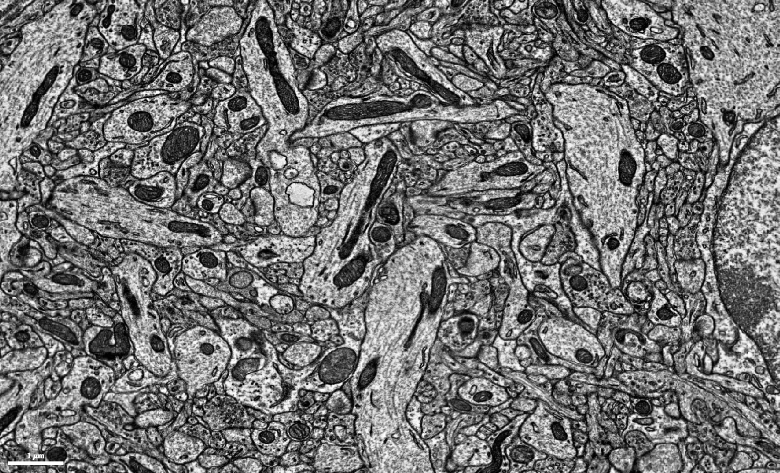
Animated sequence of serial electron microscopy sectionsSECTION FEED Z 0712 · one plane at a time
Z 1979Z 0712
VOL_07A · Z 0712
IMAGE_PASS · 1/5
SOURCE · EYEWIRE / SEUNG LAB ↗
STAGE 01

Turn tissue into voxels

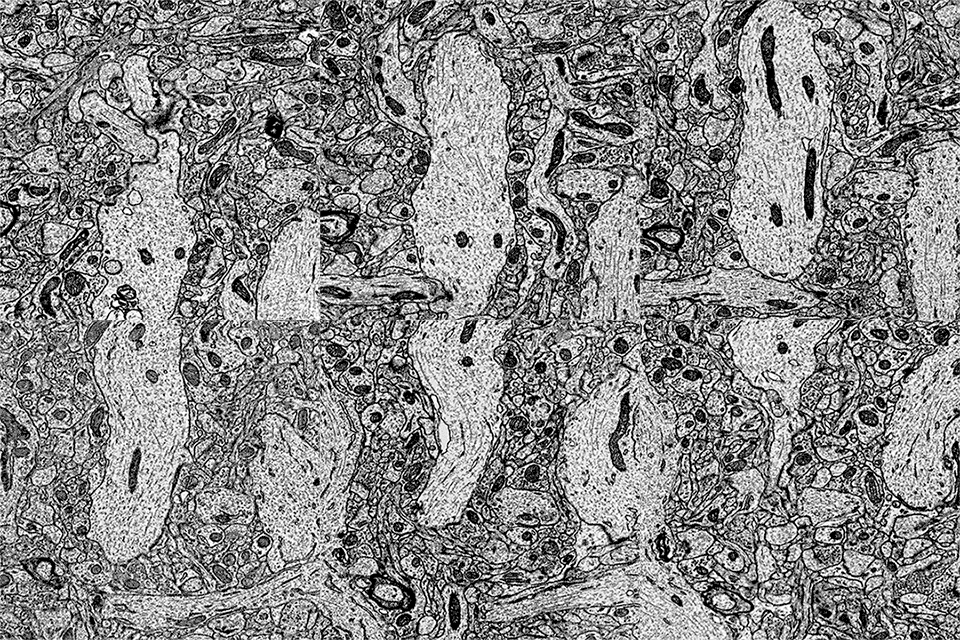
A preserved brain is stained with heavy metals, cut or milled into ultrathin layers, then scanned with an electron beam. Each image is only a flat field of membranes. Thousands of images make a volume.

4 × 4 × 30 nmtypical Fish1 voxel
02 / HUMAN IN THE LOOP

The machine draws. People decide.

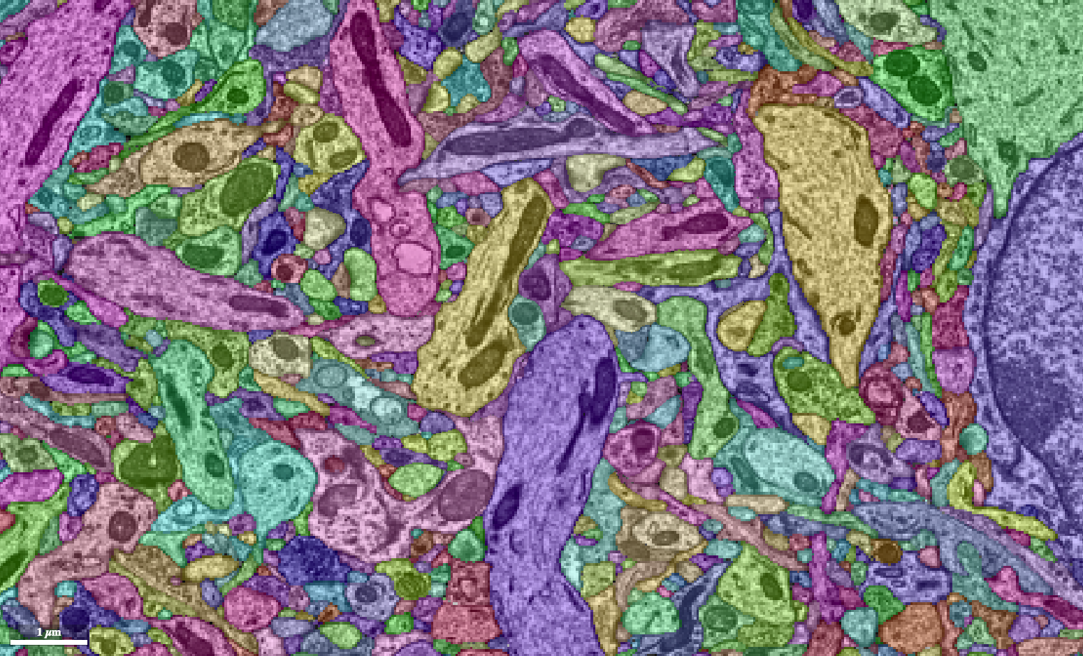
A cell is proofread through repeated branch extension and merger removal: reconnect pieces that belong to one cell, and separate branches that belong to different cells.

FALSE MERGERTwo cells share one segment.Correct it with a split.

FALSE SPLITOne cell occupies separate segments.Correct it with a merge.

PROOFREADING LAB · REPRESENTATIVE 2D + 3D VIEWS0/4 ACTIONS SAVED
Electron microscopy slice used to inspect cell boundaries
Colored segmentation aligned with the raw EM slice
2D EM · Z 344
BEFOREONE SEGMENT · TWO CELLS
3D SEGMENTATION MESH · BEFORE
TARGET / SIDE A CANDIDATE / SIDE B CUT SEEDS
1Find the false junction2Seed both sides3Preview the cut4Split the body

WHAT THE REVIEWER DOESA foreign branch has been fused to the target cell. Place red and blue seed groups on opposite sides of the false junction, preview the cut, then split the body.WATCH THE REAL WORKFLOW ↗

The EM and public MICrONS mesh render are representative views, not a co-registered case. The interactions reproduce the correction logic shown in FlyWire and NeuTu.

COLLABORATIVE LAYER

One map, many kinds of attention.

Proofreaders correct false merges and false splits, reviewers declare a reconstruction ready under project-specific criteria, and annotators attach evidence-backed labels with provenance. Researchers and citizen scientists share that careful work—and often become experts themselves.

Expert proofreader Research lab Citizen scientist
Open the EyeWire II community workspace ↗
03 / WHAT THE MAP UNLOCKS

Anatomy becomes a testable graph.

The connectome constrains what a circuit can do. Activity, molecules and behavior supply the rest of the story.

FLYWIRE CODEXFAFB · SNAPSHOT 783 · LEFT HEMISPHERE
83 SYN58 SYN50 SYN46 SYN42 SYNv2LN30LOCAL NEURON · GLUTAMATE35945919v2LN36LOCAL NEURON · SEROTONIN20541349DA1_lPNLATERAL PN · ACETYLCHOLINE03231916CB3356CENTRAL · ACETYLCHOLINE29163931DA1_vPNVENTRAL PN · GABA46122804
NODE = PROOFREAD NEURON→ EDGE = DIRECTED SYNAPSES
FAFB QUERY · PATHS

Trace a microcircuit

Five measured connections around one DA1 lateral projection neuron, returned by the official FAFB v783 connectivity example.

5 edges / 279 synapsesroot_id == 720575940603231916
FAFB v783139,255 neurons

The proofread female adult fly brain represented in Codex as a directed graph.

CODEX GRAPH3,732,460 connections

Edges aggregate synapses from one presynaptic neuron to one postsynaptic neuron.

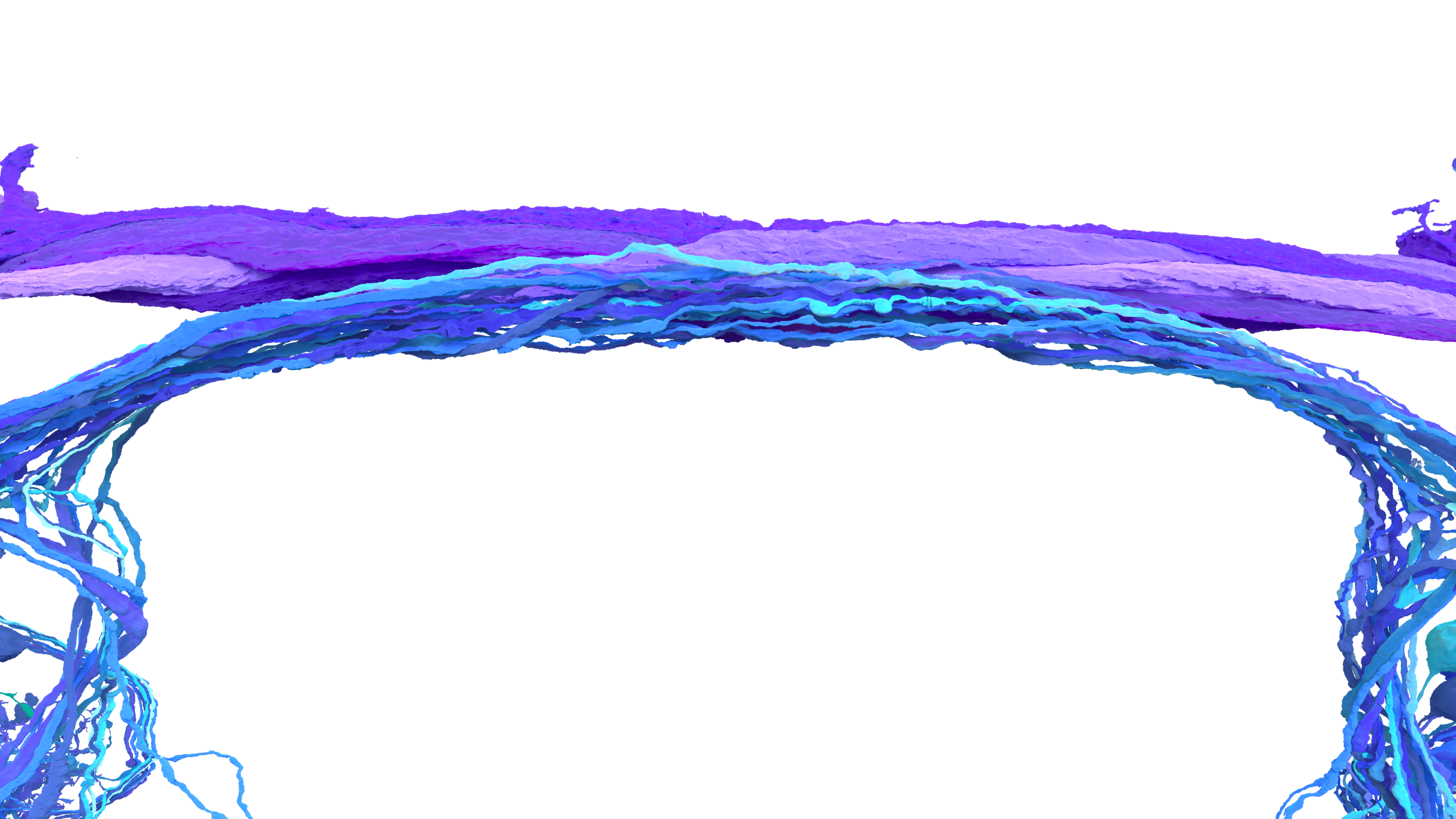
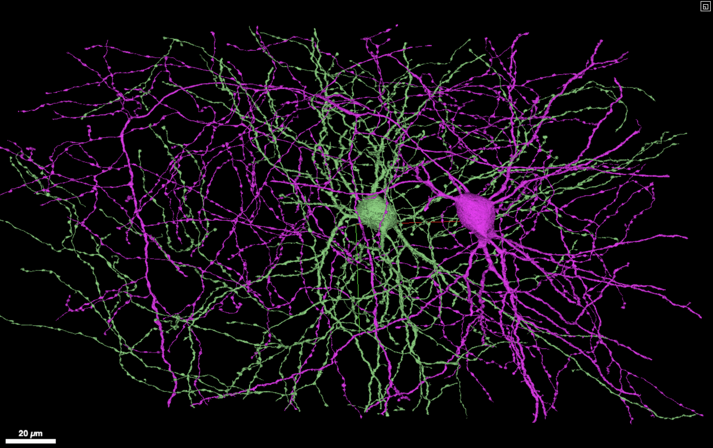
Purple and cyan reconstructed neuron cable meshes
RENDER · AMY STERLING
04 / THE NEXT FRONTIER

Questions to ask of a connectome.

A synapse-resolution reference turns the nervous system from something you observe into something you can address. You no longer sample neurons and hope. You name them.

Every capability below was impractical without a wiring diagram of named cells, and is now routine somewhere in the field. The shift is from describing a circuit to interrogating one: the map is a queryable object, and each answer it returns is a hypothesis you can take back to a live animal and try to break.

TARGET

Record the cell you meant to record

Choose which neuron to patch, image or ablate because its partners are already known. Physiology stops being a lottery over whatever the pipette found.

PREDICT

Simulate, then try to falsify

Run the measured graph forward and ask what it should do. A prediction that survives contact with real activity is evidence; one that fails localises the missing biology.

CLASSIFY

Type a cell by who it talks to

Connectivity separates cell types that no stain distinguishes, and it names the ones nobody has described yet.

TRACE

Walk backwards from behaviour

Start at a muscle and follow the graph upstream to the specific cells that drive it, then test them one at a time.

COMPARE

Ask what is conserved

Two individuals, or two species, and the same circuit in both. Difference becomes measurable rather than anecdotal.

INTERVENE

Design the experiment on the map first

Pick the minimal set of cells whose silencing should change a behaviour, before touching an animal.

What wiring still does not give you. A connectome says where signals may travel, not the rules by which each cell transforms them. Neurotransmitter identity, receptor complement, plasticity and a body that keeps changing all sit outside the graph. The target is not a frozen diagram but a versioned, multimodal model of a living circuit, and every question above gets sharper as those layers arrive.

05 / DATASET OBSERVATORY

The public connectomics field.

A living catalog of major public micro connectomics datasets. Last reviewed August 2026. For a list of macro connectomics datasets, visit this page.

23records shown
40 YEARS OF OPEN MAPS

From one worm to a world of brains.

Released datasets lead into active programs. Scroll sideways to move from the first complete nervous-system connectome to the current global frontier.

PUBLIC DATASET ACTIVE PROGRAMEvery card states its mapping scale.
  1. 1986UK + USA
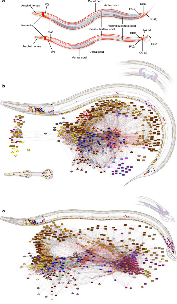
    C. elegans neuroanatomy and whole-animal connectivity diagram
    Cook et al. · Nature 2019
    SYNAPSE

    C. elegans

    The first complete nervous-system wiring diagram: 302 neurons traced by serial electron microscopy.

    OPEN SOURCE ↗
  2. 2016USA + JAPAN
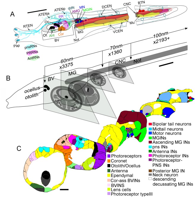
    Ciona larva nervous system reconstructed from serial electron microscopy
    Ryan, Lu & Meinertzhagen · eLife 2016
    SYNAPSE

    Ciona larva

    A complete central nervous system connectome for a tadpole larva, spanning 177 CNS neurons.

    OPEN SOURCE ↗
  3. 2020USA + GLOBAL
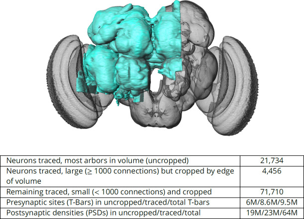
    Reconstructed hemibrain volume highlighted inside an adult Drosophila brain
    Scheffer et al. · eLife 2020
    SYNAPSE

    Drosophila hemibrain

    A densely reconstructed adult fly central brain with roughly 25,000 neurons and an open graph in neuPrint.

    OPEN SOURCE ↗
  4. 2023UK + GLOBAL
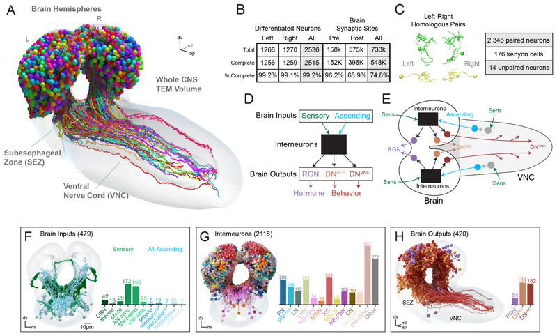
    Complete Drosophila larva brain reconstruction with neurons colored by class
    Winding et al. · Science 2023 · CC BY
    SYNAPSE

    Drosophila larva

    The first complete insect brain connectome: 3,016 neurons linked across both hemispheres.

    OPEN SOURCE ↗
  5. 2024GLOBAL
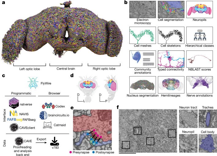
    FlyWire whole-brain reconstruction and its electron microscopy, segmentation and analysis layers
    Dorkenwald et al. · Nature 2024 · CC BY
    SYNAPSE

    FlyWire / FAFB

    The adult female fly brain became a public, queryable graph of 139,255 neurons.

    OPEN SOURCE ↗
  6. 2024USA
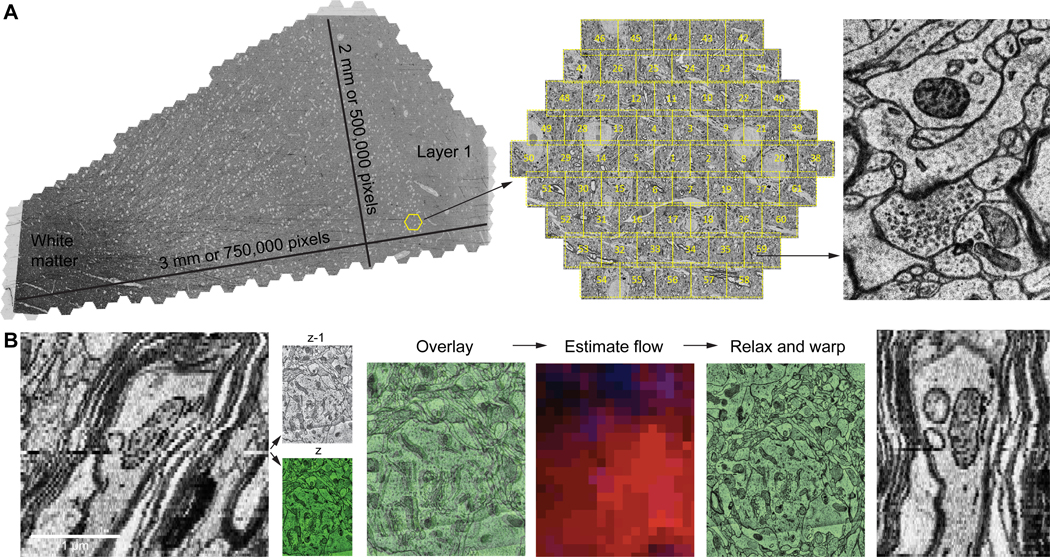
    H01 human cortex electron microscopy volume, image tiles and aligned cellular ultrastructure
    Shapson-Coe et al. · Science 2024
    SYNAPSE

    H01 human cortex

    A cubic millimeter of human temporal cortex: more than 50,000 cells in an open petascale volume.

    OPEN SOURCE ↗
  7. 2024CHINA
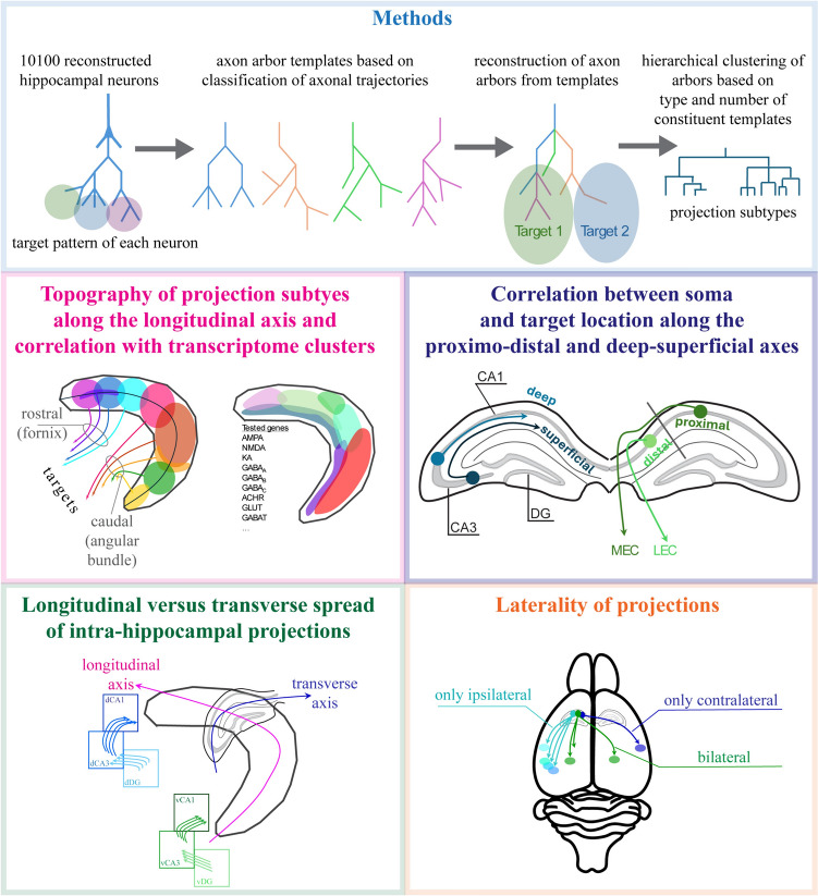
    Summary of the Digital Brain hippocampal single-neuron projectome and projection subtypes
    Qiu et al. dataset · 2024
    MESOSCALE

    Digital Brain projectome

    More than 10,000 mouse hippocampal neurons reconstructed across their whole-brain axonal projections.

    OPEN SOURCE ↗
  8. 2025USA + GLOBAL
    MICrONS neurons reconstructed within a cubic millimeter of mouse visual cortex
    MICrONS Explorer · Allen Institute
    SYNAPSE + FUNCTION

    MICrONS cortical mm³

    A cubic millimeter of mouse visual cortex joins roughly 200,000 cells and half a billion synapses to activity.

    OPEN SOURCE ↗
  9. 2023 →USA
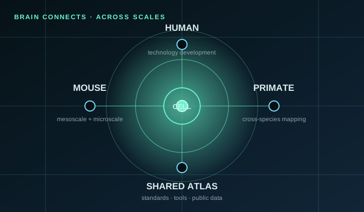
    BRAIN CONNECTS diagram linking mouse, primate and human mapping across scales
    Diagram: Connectome Atlas · source: NIH BRAIN
    MULTISCALE

    BRAIN CONNECTS

    An NIH network developing technologies and shared infrastructure for whole-brain wiring diagrams in mouse, primate and human.

    ACTIVE PROGRAM ↗
  10. ACTIVEJAPAN
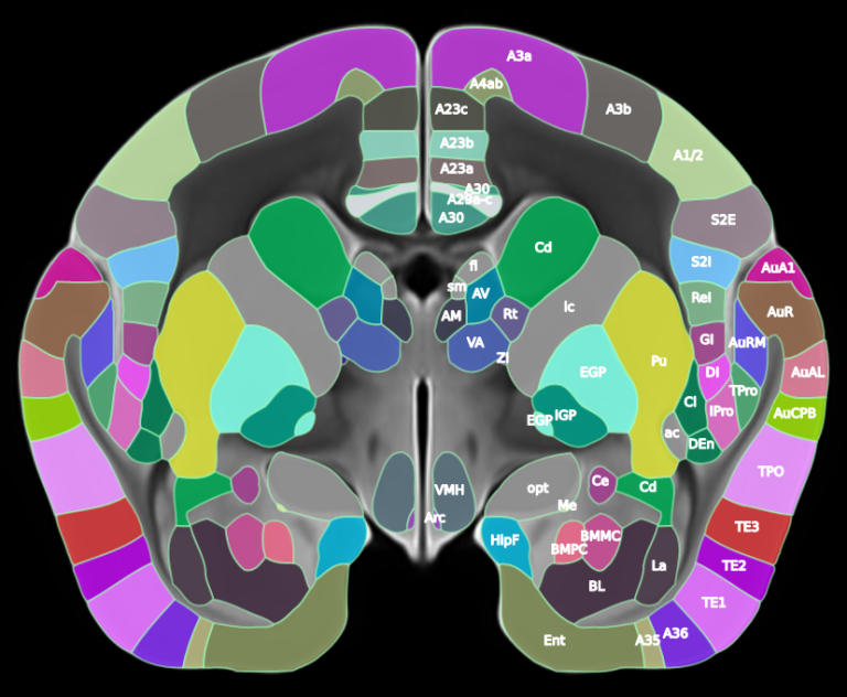
    Brain/MINDS marmoset brain atlas with labeled cortical and subcortical regions
    Brain/MINDS Data Portal · RIKEN
    MESOSCALE + MULTIMODAL

    Brain/MINDS 2.0

    Marmoset and human mapping integrates neuronal tracing, gene expression and imaging into public cross-species atlases.

    ACTIVE PROGRAM ↗
  11. ACTIVECHINA + GLOBAL
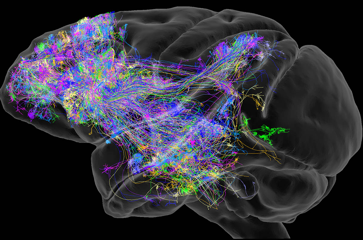
    Dense colored projection pathways rendered inside a primate brain
    Chinese Academy of Sciences · 2025
    MESOSCALE

    International primate brain atlas

    A China-led collaboration is building cross-species mesoscale resources toward primate and human wiring atlases.

    ACTIVE PROGRAM ↗
  12. ACTIVEEUROPE
    EBRAINS human reference atlas with cortical regions rendered in distinct colors
    EBRAINS · siibra reference atlas
    MULTISCALE

    EBRAINS 2.0

    Living human, macaque and rodent atlases continue to integrate anatomy, connectivity and function across scales.

    ACTIVE PROGRAM ↗

Scale note. Synapse-resolution connectomes, single-neuron projectomes and multiscale brain atlases answer different questions. The timeline keeps them together to show the field’s trajectory—not to imply that they are technically equivalent.

Name / organismCoverageCellsReleasedAccess
C. elegans adult hermaphroditeNematodeEntire animalcomplete3021986WormWiring ↗C. elegans adult maleNematodeEntire animalcomplete3852019WormWiring ↗C. elegans development seriesNematodeEntire animal · 8 agescomplete300–3022021WormWiring ↗Ciona larvaSea squirtCentral nervous systemcomplete177 CNS2016ANISEED / paper ↗Platynereis 3-day larvaMarine annelidEntire animalcomplete~2,7002025CATMAID ↗Drosophila larvaFruit flyBrain + nerve cordcomplete3,0162023CATMAID ↗FlyWire / FAFBFruit flyAdult female braincomplete139,2552024Codex · FlyWire ↗BANCFruit flyAdult female brain + VNCcomplete~160K2025CAVE · Codex ↗Male CNSFruit flyAdult male brain + VNCcomplete~170K2025neuPrint ↗Hemibrain v1.2.1Fruit flyAdult central braindense volume~25K2020neuPrint ↗MANCFruit flyAdult male nerve corddense volume~23K2023neuPrint ↗FANCFruit flyAdult female nerve corddense volume~14K2024CAVE ↗Mushroom bodyFruit flyLearning + memory centerdense volume~2K2017neuPrint ↗Medulla 7-columnFruit flyOptic lobedense volume~1K2015neuPrint ↗Fish1ZebrafishLarval brain + anterior cordin progress187,053 somata2025CAVE · Neuroglancer ↗MICrONS mm³MouseVisual cortexdense volume200K cells2025CAVE · Neuroglancer ↗MICrONS phase 1MouseVisual cortex L2/3dense volume364 excitatory2022CAVE · Neuroglancer ↗EyeWire e2198MouseRetinapartial~1K traced2014EyeWire ↗EyeWire IIMouse1 mm² retinain progress>25K complete2026EyeWire II ↗H01HumanTemporal cortex · 1 mm³dense volume>50K cells2024Neuroglancer ↗Kasthuri15MouseSomatosensory cortexdense volume1,700 fragments2015BossDB ↗Pinky40MouseVisual cortexdense volumedense segmentation2019BossDB · CAVE ↗Hippocampus CA3MouseCA3 pyramidal layerpartial38 proofread2019CAVE ↗

Scope note. “Complete” means complete to the authors’ declared anatomical and technical scope, not error-free or biologically exhaustive. Connectomes remain versioned scientific objects.

Suggest a missing dataset ↗
06 / SOURCE SIGNALS

Keep exploring.

07 / PUBLIC LAB

Enter a living connectome.

This is the EyeWire II community branch of Neuroglancer: real EM, real segmentation and a path into public participation in connectomics.

LIVE EXTERNAL WORKSPACEMINNIE · PINKY · XY + 3DOPEN FULL SCREEN ↗
COMMUNITY-RUN SCIENCE

The Atlas does not control this external workspace. Viewing is public; contributing edits may require connecting to the EyeWire II community system.

READ THE COMMUNITY GUIDE ↗