The five visible edge weights are the first five rows in the official fafbseg v783 connectivity example for root 720575940603231916. They are individual directed neuron-pair weights, summed only for the small educational graph.
Inspect basis ↗EVIDENCE LEDGER / REVIEWED AUGUST 2026
Receipts for the map.
Sources, calculations, and credits for the Connectome Atlas.
32 CATALOG RECORDS15 CORE PAPERS11 CREDIT LINES
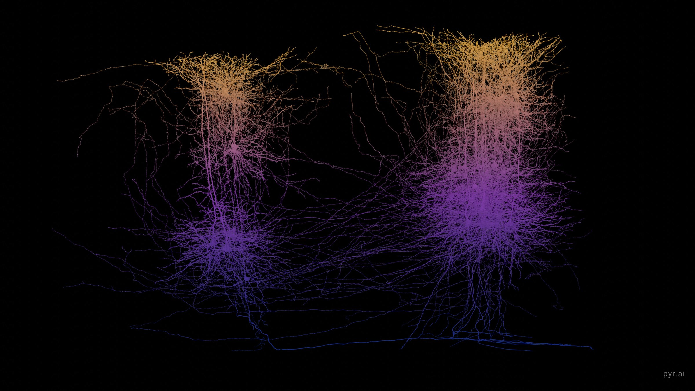
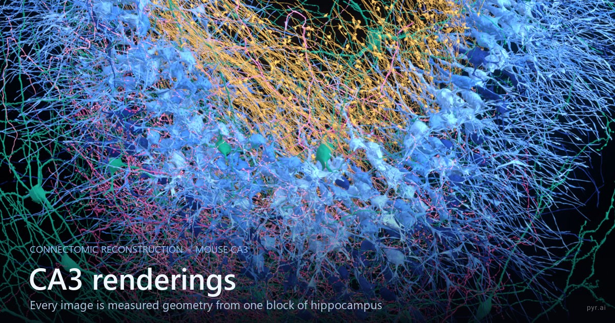
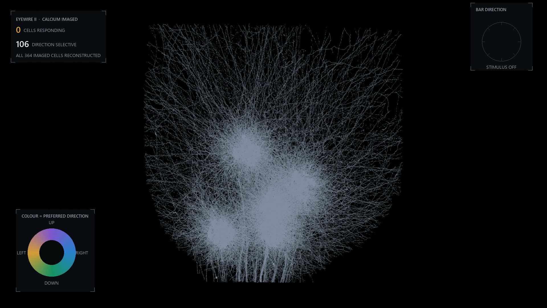
SELECTED RENDERS / AMY STERLING
The atlas in pictures.
Connectomics renders by Amy Sterling, made from public reconstruction data.
01 / PRIMARY LITERATURE
Core scientific references.
Primary papers behind the atlas, grouped by research area.
015 REFERENCES
FW01FlyWire reconstructionFlyWire & FAFB
Whole-brain reconstruction, annotation, analysis and community proofreading
Dorkenwald, S. et al. Neuronal wiring diagram of an adult brain. Nature 634, 124–138 (2024).
10.1038/s41586-024-07558-y↗FW02FlyWire annotationsSchlegel, P. et al. Whole-brain annotation and multi-connectome cell typing of Drosophila. Nature 634, 139–152 (2024).
10.1038/s41586-024-07686-5↗FW03FlyWire graph analysisLin, A., Yang, R. et al. Network statistics of the whole-brain connectome of Drosophila. Nature 634, 153–165 (2024).
10.1038/s41586-024-07968-y↗FW04FAFB connectivity dataFlyWire Consortium. FlyWire Whole-brain Connectome Connectivity Data, release 783.0. Zenodo (2024).
10.5281/zenodo.10676866↗FW05Community proofreadingDorkenwald, S. et al. FlyWire: online community for whole-brain connectomics. Nature Methods 19, 119–128 (2022).
10.1038/s41592-021-01330-0↗022 REFERENCES
PF013D proofreading operationsProofreading infrastructure
3D merge/split operations, collaboration, edit history and versioned annotations
Zhao, T. et al. NeuTu: Software for Collaborative, Large-Scale, Segmentation-Based Connectome Reconstruction. Frontiers in Neural Circuits 12, 101 (2018).
10.3389/fncir.2018.00101↗PF02Versioned proofreading & annotationDorkenwald, S. et al. CAVE: Connectome Annotation Versioning Engine. Nature Methods 22, 1112–1120 (2025).
10.1038/s41592-024-02426-z↗034 REFERENCES
CE01C. elegansWhole-animal connectomes
Foundational nervous-system maps across model organisms
White, J. G., Southgate, E., Thomson, J. N. & Brenner, S. The structure of the nervous system of the nematode Caenorhabditis elegans. Phil. Trans. R. Soc. B 314, 1–340 (1986).
10.1098/rstb.1986.0056↗CE02C. elegans, both sexesCook, S. J. et al. Whole-animal connectomes of both Caenorhabditis elegans sexes. Nature 571, 63–71 (2019).
10.1038/s41586-019-1352-7↗CI01Ciona larvaRyan, K., Lu, Z. & Meinertzhagen, I. A. The CNS connectome of a tadpole larva of Ciona intestinalis. eLife 5, e16962 (2016).
10.7554/eLife.16962↗DL01Drosophila larvaWinding, M. et al. The connectome of an insect brain. Science 379, eadd9330 (2023).
10.1126/science.add9330↗042 REFERENCES
MI01MICrONSMammalian cortex
Synapse-resolution mouse and human cortical volumes
MICrONS Consortium. Functional connectomics spanning multiple areas of mouse visual cortex. Nature 640, 435–447 (2025).
10.1038/s41586-025-08790-w↗H001Human cortex / H01Shapson-Coe, A. et al. A petavoxel fragment of human cerebral cortex reconstructed at nanoscale resolution. Science 384, eadk4858 (2024).
10.1126/science.adk4858↗052 REFERENCES
NM01Field overviewField methods & modelling
Connectomics methods and embodied circuit simulation
Marx, V. Method of the Year: EM connectomics. Nature Methods 22, 2470–2475 (2025).
10.1038/s41592-025-02906-w↗SIM1Embodied modellingLobato-Rios, V. et al. NeuroMechFly, a neuromechanical model of adult Drosophila melanogaster. Nature Methods 19, 620–627 (2022).
10.1038/s41592-022-01466-7↗02 / DATA SOURCE REGISTRY
Where each catalog record points.
Project portals are used when a resource is living or versioned; DOI papers are used for fixed publication releases.
Dataset familyCanonical sourceFigure shownLink
C. elegans adult + developmentWormWiring302 / 385 neurons; developmental seriesOPEN ↗Ciona larvaeLife data and ANISEED177 CNS neuronsOPEN ↗Platynereis larvaJékely Lab / published whole-animal resource~2,700 cells in the catalog recordOPEN ↗Drosophila larvaWinding et al. / CATMAID3,016 neuronsOPEN ↗FlyWire / FAFBFlyWire Codex v783139,255 neurons; 3,732,460 Codex connectionsOPEN ↗BANCFlyWire CodexVersioned female brain + VNC releaseOPEN ↗Hemibrain, MANC, Male CNSJanelia FlyEM / neuPrintPublished partial or CNS connectomesOPEN ↗FANCFANC communityFemale adult nerve cordOPEN ↗Fish1Fish1 public releaseLarval zebrafish brain + anterior cordOPEN ↗MICrONS mm³ + phase 1MICrONS ExplorerMouse visual cortex structure × functionOPEN ↗EyeWire retina + EyeWire IIEyeWire / Seung Lab communityMouse retinal reconstructions and public proofreadingOPEN ↗H01Google Research / Lichtman LabHuman temporal cortex sampleOPEN ↗Kasthuri15 + Pinky40BossDBMouse cortical EM volumesOPEN ↗Hippocampus CA3Public CA3 viewer38 proofread cells in this catalog recordOPEN ↗Macro catalogHCP, UK Biobank, ABCD, dHCP, Allen, marmoset and CoCoMac portalsNine region-scale records, each linked to its owning projectOPEN ↗Version rule. “Last reviewed” means the catalog links and displayed figures were checked in August 2026. It does not freeze the linked portal. Before quoting a number, cite the project’s named release or materialization.
03 / IMAGE, SOFTWARE & COMMUNITY CREDITS
The map has many makers.
01Created by
02Interface system
amyleesterling/scifi-ui, adapted for the Connectome Atlas
03Hero and social preview
Connectome rendering supplied by Amy Sterling / What Is a Brain?
04Serial EM and segmentation teaching media
EyeWire / Seung Lab educational materials; each pipeline view links to its source
05MICrONS viewer imagery
MICrONS Explorer and its public Neuroglancer scenes
06Dataset timeline figures
Cook et al.; Ryan, Lu & Meinertzhagen; Scheffer et al.; Winding et al.; Dorkenwald et al.; Shapson-Coe et al.; Qiu et al.; and the MICrONS Explorer
07Program timeline imagery
NIH BRAIN CONNECTS (diagram adapted by Connectome Atlas), Brain/MINDS Data Portal / RIKEN, Chinese Academy of Sciences, and EBRAINS / siibra
08Nature Methods December 2025 cover
Image: Amy Sterling, FlyWire and Princeton University. Cover design: Thomas Phillips. © Springer Nature
09FAFB reconstruction and annotations
FlyWire Consortium, Princeton Neuroscience Institute, Cambridge/MRC LMB researchers and the FlyWire community
10Volume-viewing software
Neuroglancer, Google Research
11Public proofreading context
EyeWire and EyeWire II communities
CORRECTIONS WELCOME
This is a living scholarly object. If a count, citation, release label or credit is incomplete, send the exact correction and its source.
Submit a correction ↗04 / CALCULATIONS & DEFINITIONS
How the displayed numbers were made.
Reproducible arithmetic where possible; plain disclosure where the interface is illustrative.
Codex counts qualifying directed neuron-to-neuron edges in its FAFB graph. The FlyWire paper reports individual chemical synapses. Many synapses can contribute to one edge; Codex uses a default minimum of five synapses for FAFB connections.
Inspect basis ↗This is a count of hand-curated records displayed by this site, not a scientific estimate of every connectomics dataset ever produced. Subvolumes and successive releases can overlap.
Inspect basis ↗The z number, registration confidence, pass counters and proofread score in the pipeline are explanatory interface simulations. They are not measurements derived from the shown microscopy images.
Inspect basis ↗